Simple Compact Bone Diagram / Osteon Wikipedia - Compact bone definition compact bone, also known as cortical bone, is a denser material used to create much of the hard structure of the skeleton.
Simple Compact Bone Diagram / Osteon Wikipedia - Compact bone definition compact bone, also known as cortical bone, is a denser material used to create much of the hard structure of the skeleton.. The remainder is cancellous bone, which has a spongelike appearance with numerous large spaces and is found in the. Osteons are the small units of which the hardest parts of human bones are made. In compact bone, these cells are embedded within the solid calcium phosphate matrix of solid bone. The main type of bone cell is the osteocyte (bone cell, shown as purple in the diagram). Anatomy of shoulder 12 photos of the anatomy of shoulder anatomy of nerves in shoulder, anatomy of posterior shoulder dislocation, anatomy of right shoulder, anatomy of shoulder labrum tear, anatomy of the shoulder games, human anatomy, anatomy of nerves in shoulder, anatomy of posterior shoulder dislocation, anatomy of.
Learn about the process of bone formation. Recorded at glen oaks community college, centreville, michigan by dr ren allen hartung. It makes up the outer cortex of all bones and is in immediate contact with the periosteum. Related posts of compact bone diagram labeled anatomy of shoulder. Compact bone diagram compact bone diagram spongy bone diagram the histology guide cartilage bone ossification.
Spongy Bone Histology Microanatomy Web Atlas Gwen V Childs Ph D from microanatomy.net Bone formation, process by which new bone is produced. This video describes the microscopic anatomy of compact bone. The diagram above shows a longitudinal view of an osteon. Compact bone is the denser, stronger of the two types of osseous tissue (figure 6.3.6). Cortical bone forms a dense cylinder down the shaft of the bone surrounding the central marrow cavity. The process takes two general forms, one for compact bone and the other for cancellous bone. It makes up the outer cortex of all bones and is in immediate contact with the periosteum. Animal cell structures functions diagrams simple animal cell drawing at getdrawingscom free for personal 50 diagram of the cell noibaiairporttransfer
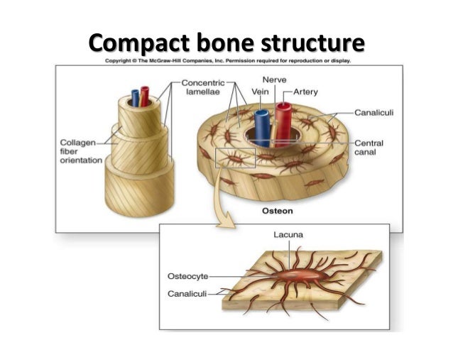
(b) in this micrograph of the osteon, you can clearly see the concentric lamellae and central canals.
This provides the bones strength and consists of tightly stacked layers of bone which appear to form a solid section. Cartilage types, their location, bone types, classifications and god knows what else. The compact bones form the hard exterior of the bones, whereas the spongy bones have several pores that are filled with nerves and blood vessels. Flussdiagramm, mind map, prozessdiagramm, organigramme, uml, er usw. Some, mostly older, compact bone is remodelled to form these haversian systems (or osteons). Deep to the compact bone layer is a region of spongy bone where the bone tissue grows in thin columns called. Compact bone definition compact bone, also known as cortical bone, is a denser material used to create much of the hard structure of the skeleton. Under the periosteum is a thin layer of compact bone (often called cortical bone). Diagram of a typical long bone: Bone formation, process by which new bone is produced. The remainder of the bone is formed by cancellous or spongy bone. Diagram of distinct morphological types of bone. Despite the fact that the soft bone tissue is softer than compact bone tissue, the vascular activity is quite high and its unique design gives bones a considerable boost in strength.
Ossification begins about the third month of fetal life in humans and is completed by late adolescence. The compact bones form the hard exterior of the bones, whereas the spongy bones have several pores that are filled with nerves and blood vessels. Compact bone is made of a matrix of hard mineral salts reinforced with tough collagen fibers. Simple bone diagrams to help students identify and label compact bone, spongy bone, yellow marrow, periosteum, and cartilage. Terms in this set (8) spongy bone (contains red marrow) compact bone (has osteons) osteon.
Biopolymers As Bone Substitutes A Review Biomaterials Science Rsc Publishing from pubs.rsc.org (b) in this micrograph of the osteon, you can clearly see the concentric lamellae and central canals. Each cell appears to be isolated from other cells, but in reality are connected to neighboring cells by thin cellular extensions that pass through tiny channels in the solid matrix (see picture on left of. These are shown in the figure below. The process takes two general forms, one for compact bone and the other for cancellous bone. (b) in this micrograph of the osteon, you can clearly see the concentric lamellae and central canals. Some, mostly older, compact bone is remodelled to form these haversian systems (or osteons). They are roughly cylindrical, and about 0.2mm wide and a few millimeters long. The remainder of the bone is formed by cancellous or spongy bone.
Compact bone is the denser, stronger of the two types of osseous tissue (figure 6.3.6).
Simple bone diagrams to help students identify and label compact bone, spongy bone, yellow marrow, periosteum, and cartilage. Osteons are the small units of which the hardest parts of human bones are made. Some, mostly older, compact bone is remodelled to form these haversian systems (or osteons). Shows compact (cortical) and cancellous (spongy) bone. Terms in this set (8) spongy bone (contains red marrow) compact bone (has osteons) osteon. Learn about the process of bone formation. Compact bone is made of a matrix of hard mineral salts reinforced with tough collagen fibers. Compact bone is the denser, stronger of the two types of osseous tissue (figure 6.3.6). Flussdiagramm, mind map, prozessdiagramm, organigramme, uml, er usw. Create flashcards for free and quiz yourself with an interactive flipper. (b) in this micrograph of the osteon, you can clearly see the concentric lamellae and central canals. Many tiny cells called osteocytes live in small spaces in the matrix and help to maintain the strength and integrity of the compact bone. Long bones such as the femur contain two distinct morphological types of bone:
Compact bone, also called cortical bone, dense bone in which the bony matrix is solidly filled with organic ground substance and inorganic salts, leaving only tiny spaces (lacunae) that contain the osteocytes, or bone cells.compact bone makes up 80 percent of the human skeleton; Flussdiagramm, mind map, prozessdiagramm, organigramme, uml, er usw. Compact bone is the denser, stronger of the two types of osseous tissue (figure 6.3.6). Cardiac nursing pediatric nursing structure of bone anatomy bones anatomy art types of bones medical massage medical pictures bones. They are roughly cylindrical, and about 0.2mm wide and a few millimeters long.
Histo Bone from image.slidesharecdn.com Each cell appears to be isolated from other cells, but in reality are connected to neighboring cells by thin cellular extensions that pass through tiny channels in the solid matrix (see picture on left of. Compact bone is made of a matrix of hard mineral salts reinforced with tough collagen fibers. Cortical bone forms a dense cylinder down the shaft of the bone surrounding the central marrow cavity. Compact bone, also called cortical bone, dense bone in which the bony matrix is solidly filled with organic ground substance and inorganic salts, leaving only tiny spaces (lacunae) that contain the osteocytes, or bone cells.compact bone makes up 80 percent of the human skeleton; Despite the fact that the soft bone tissue is softer than compact bone tissue, the vascular activity is quite high and its unique design gives bones a considerable boost in strength. The remainder is cancellous bone, which has a spongelike appearance with numerous large spaces and is found in the. Create flashcards for free and quiz yourself with an interactive flipper. Animal cell structures functions diagrams simple animal cell drawing at getdrawingscom free for personal 50 diagram of the cell noibaiairporttransfer
Shows compact (cortical) and cancellous (spongy) bone.
Cortical bone forms a dense cylinder down the shaft of the bone surrounding the central marrow cavity. Article by jennifer smith owens. Long bones such as the femur contain two distinct morphological types of bone: Learn about the process of bone formation. (b) in this micrograph of the osteon, you can clearly see the concentric lamellae and central canals. Compact bone definition compact bone, also known as cortical bone, is a denser material used to create much of the hard structure of the skeleton. Many tiny cells called osteocytes live in small spaces in the matrix and help to maintain the strength and integrity of the compact bone. It is also called osseous tissue or cortical bone and it provides structure and support for an organism as part of its skeleton, in addition to being a location for the storage of minerals like calcium.about 80% of the weight of the human skeleton comes from. In long bones, as you move from the outer cortical compact bone to the inner medullary cavity, the bone transitions to spongy bone. The remainder of the bone is formed by cancellous or spongy bone. (b) in this micrograph of the osteon, you can clearly see the concentric lamellae and central canals. Diagram of a typical long bone: Under the periosteum is a thin layer of compact bone (often called cortical bone).
Learn about the process of bone formation compact bone diagram. Shows compact (cortical) and cancellous (spongy) bone.


0 Komentar